Jesse Gomez
Graduate Student
The anatomical, functional, and behavioral development of high-level vision
What aspects of visual cortex change across development as children improve at reading and recognizing everyday objects?
Do changes in brain function necessitate changes in structure? What happens when this development goes awry?
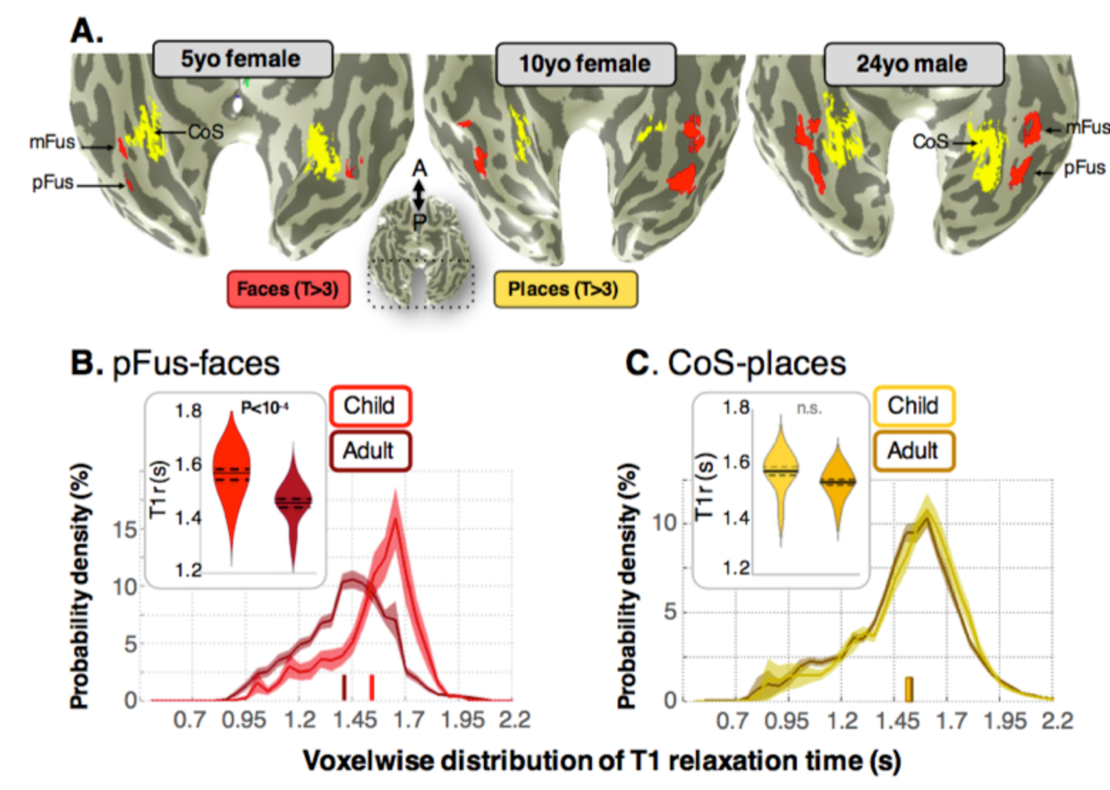
To answer some of these questions, we have undertaken a longitudinal research study of human high-level visual cortex
in children as young as five years old. We find that there is growth of tissue and within portions of ventral temporal cortex,
challenging the idea that cortex is either quiescent or pruned after early childhood. This tissue growth, within
face-selective cortex as an example, is correlated with increases in the functional selectivity of face-selective regions
and linked to improvements in recognition memory ability well into adulthood (Gomez et al., 2016 in revision).
This novel observation was made possible by combining new quantitative magnetic resonance imaging (qMRI) techniques
with functional MRI and behavioral measurements in single-subject analyses. We have further linked these in vivo
anatomical measurements with ex vivo quantifications of cytoarchitecture in a collaboration with the
Institute of Neuroscience and Medicine in Julich, Germany (INM-1).
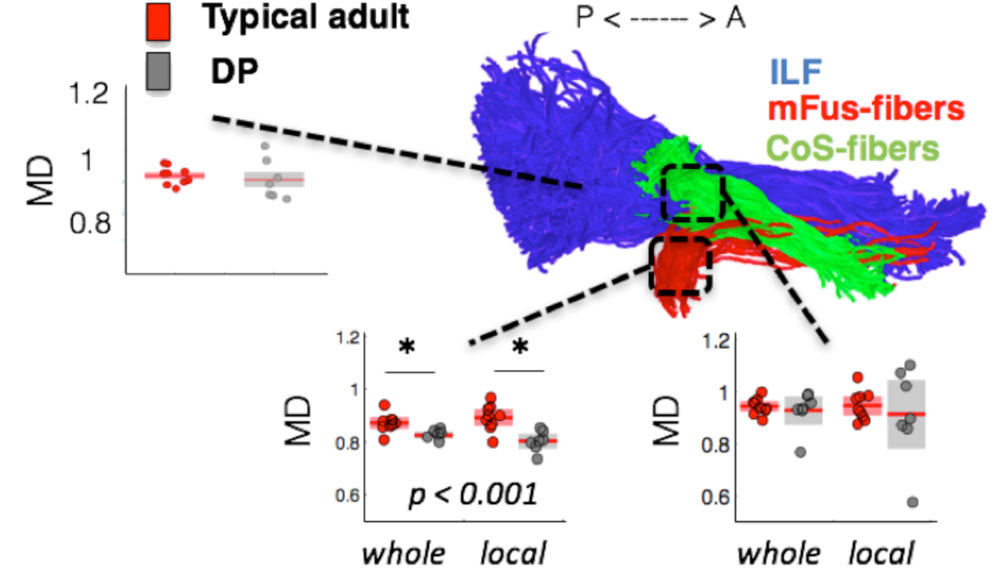
In earlier work, we demonstrated that the organization of high-level visual cortex may be constrained by
parallel white matter tracts corresponding to functional divisions in the cortex, and that anatomical properties
of these tracts are behaviorally relevant, as they differentiate adults with a face recognition deficit known as
developmental prosopagnosia (Gomez et al., 2015 Neuron).
Future work will aim to measure changes in population
receptive fields in order to answer how pooling of visual information from early visual field maps develops into adulthood.
|
|
|